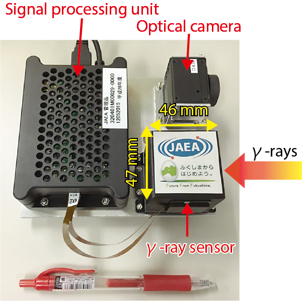
Fig.1-16 Photograph of a compact Compton camera
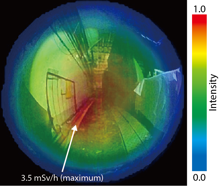
Fig.1-17 Visualization result of radioactive substances inside 1F3’s turbine building
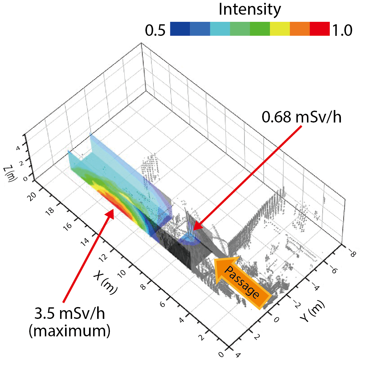
Fig.1-18 3D-radiation-distribution map
Seeing invisible radiation is important not only for protecting workers from radiation exposure, but also for understanding the contamination situation inside the TEPCO’s Fukushima Daiichi NPS (1F). Inside its buildings, radioactive substances are adhering to various objects such as floors, walls, equipment, and scattered rubble, and the contamination due to radioactive substances exists in three dimensions (3D). There is also an area with such a high dose rate that workers cannot enter or remain there. Drawing a radiation-distribution map indicating the distribution condition of radioactive substances inside 1F is extremely important for predicting the risk to workers and decreasing the amount of radiation exposure for performing decommissioning tasks. Such a map would help workers to easily recognize the locations of radioactive substances in the work environment. In addition, the information would be effective for establishing detailed decontamination plans. We have newly developed a method for visualizing the radioactive substances scattered inside the 1F building in 3D.
To this end, we fabricated a compact Compton camera that could be installed in remote equipment such as a drone or crawler robot, as shown in Fig.1-16. This camera weighs about 680 g and was fabricated based on handheld-Compton-camera technology jointly developed by Waseda University and Hamamatsu Photonics. In high-dose-rate environments, it was necessary to equip the γ-ray sensor with a large and heavy shield for radiation measurements using a conventional Compton camera. On the other hand, since our Compton camera is compact, it is possible to perform measurements with a compact shield while maintaining portability within the 1F buildings. Using this Compton camera, we conducted a measurement of the distribution of radioactive substances inside 1F3’s turbine building. We succeeded in detecting a high-dose-rate region (up to 3.5 mSv/h) inside the turbine building with an air dose rate of 0.4 to 0.5 mSv/h. Fig.1-17 shows the result of the radiation measurement, which visualizes the radioactive substances (mainly 137Cs) by superimposing their image onto a photograph of the measurement environment. The measurement time required for visualization was under 1 minute.
In addition, we also succeeded in drawing a 3D-radiation-distribution map by superimposing the image of the radioactive substances obtained by the Compton camera onto a 3D-structural model acquired using a scanning-laser range finder, as shown in Fig.1-18. Two contaminated regions with a higher dose rate than the surroundings are displayed at the wall and floor surfaces of the measurement environment. Since this map reflects the dimensions and appearance of the actual work environment, workers can easily recognize dangerous regions at a glance. We believe that it will be useful for reducing worker exposure and planning decontamination.
We are currently installing this system on a small crawler robot, and we are also developing a radiation-imaging system to understand the contamination situation inside the 1F reactor buildings.

