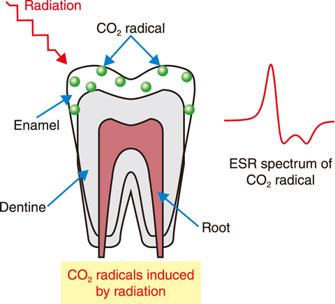
Fig.4-9 CO2 radicals induced by radiation
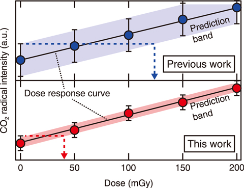
Fig.4-10 Dose–response curve for tooth enamel
The release of radioactive materials from the TEPCO’s Fukushima Daiichi NPS (1F) to the environment has resulted in long-term low-dose (below 100 mGy) exposure to humans and animals. To understand the biological effects and ensure radiation safety for individuals, the precise estimation of exposure dose is important.
Electron spin resonance (ESR) dosimetry is a powerful tool for exposure dose estimation. During ESR dosimetry, the CO2 radical, which is induced in tooth enamel by radiation and retained with a lifetime of more than 107 years, is measured, as shown in Fig.4-9. The relationship between exposure dose and CO2 radical intensity, i.e., the dose–response curve, enables the estimation of the external exposure dose of individuals. This technique has been commonly applied to estimate high-dose exposure (from a few hundred mGy to a few Gy) in humans, for example, after exposure by an atomic bomb, the nuclear plant accident in Chernobyl, or working in the nuclear industry workers. ESR dosimetry using human teeth has been reported to have a detection limit of 100–200 mGy. However, to apply ESR dosimetry for the dose estimation due to the 1F accident, this limit must be improved. This work therefore aimed to improve the detection limit to detect the exposure dose below 100 mGy using enamel separation technique by centrifugation; the developed methodology was then applied to detect the exposure dose of wild Japanese macaque from Fukushima Prefecture.
Organic materials in dentine are known to interfere when using ESR to measure the CO2 radical; it has been necessary to remove dentine and prepare dentine-free enamel samples. However, because of the small size of macaque teeth, it is difficult to grind away with a dental bur. Here, dentine was removed based on the difference in the density between enamel and dentine. To do so, the molar teeth of Japanese macaque were crushed into grains using a cryo-press; the enamel (density of 2.0–2.1 g/cm3) was separated from dentine (density of 2.8–3.0 g/cm3) by centrifugation. By this developed methodology, we can obtain “pure” enamel than the prior one.
To obtain the dose–response curve, enamel samples were irradiated by a 60Co gamma ray up to 200 mGy (50, 100, 150, and 200 mGy) by cumulative irradiation. Before the first irradiation and after each irradiation, samples were measured by ESR to obtain a precise dose–response curve (see Fig.4-10). From this dose–response curve, the detection limit was estimated to be < 40 mGy. Seven wild Japanese macaques captured in Namie in the Fukushima Prefecture, about 15 km away from 1F, were then subjected to external dose estimation. Three of them had an estimated exposure dose between 45–81 mGy, which cannot be measured by prior methods.
The improved detection limit will enable the examination of the relationship between external radiation doses (≥ 40 mGy) and the resulting biological effects on wild Japanese macaques, as well as on Japanese field mice and raccoons in Fukushima Prefecture; future aims include assessing the exposure radiation dose for children in Fukushima Prefecture.
This study was conducted on “Dose assessment of external radiation exposure in Fukushima children using deciduous teeth”, supported by the Japan Society for the Promotion of Science (JSPS) KAKENHI Grant-in-Aid for Scientific Research (C) (No.18K09906).
(Toshitaka Oka)

