| (a) | (b) |
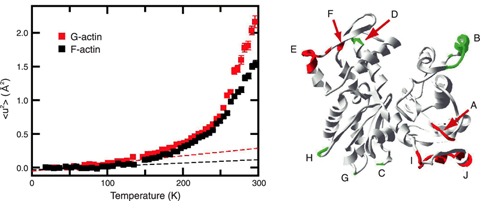
Fig.4-20
| (c) | (d) Water molecules around a protein |
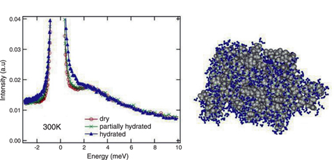
Fig.4-21
A living cell has various activities, the main players in which are proteins. The proteins are continuously exposed to thermal fluctuations of surrounding molecules, particularly water molecules which are most abundant, and the proteins themselves fluctuate. It is now well accepted that such thermal fluctuations (or dynamics) are important for the proteins to function. Complete understanding of protein function thus requires understanding of the dynamics of proteins with the water molecules around the protein.
Neutron inelastic scattering is the only method to directly "watch" thermal fluctuations of the proteins. We have been studying the dynamics of the proteins and the effects of the water molecules on them using various techniques of neutron inelastic scattering. Fig.4-20 shows the temperature dependence of the mean square displacements (MSD) of the protein actin, estimated from the neutron inelastic scattering experiments. The MSD is a direct measure of the amplitude of the fluctuations of the proteins. Actin has distinct structural states: the monomeric state (G-actin) and the polymerized state (F-actin). It was shown for the first time that G-actin and F-actin have different amplitudes of fluctuations. Furthermore, it was suggested that such differences arise from the different behavior of the loop regions involved with polymerization on the surface of the actin molecule. These results provide important information for elucidating the polymerization mechanism of actin.
How are such dynamics of the proteins affected by the surrounding water molecules? To investigate the effects of the water molecules, we performed neutron inelastic scattering experiments on the protein Staphylococcal nuclease (SNase), at different degrees of hydration. Fig.4-21 shows neutron inelastic scattering spectra of SNase in the dry state, the fully hydrated state (the protein is fully covered with a layer of water molecules), and a partially hydrated state. The spectra in the dry state and the partially hydrated state are similar, while the spectrum in the fully hydrated state shows inelastic scattering which indicates large fluctuations. This indicates that water molecules surrounding the protein are indispensable for the large fluctuations of the protein to occur, and thus are indispensable for the proteins to function.